
Tissue Section Immunostaining
Immunostaining plays a pivotal role in biomedical research. Immunohistochemistry (IHC) and immunofluorescence (IF) are among the most widely used techniques. Prior to staining, tissues or cells are fixed to preserve cellular morphology and tissue architecture; for IHC, antigen retrieval is performed as appropriate. Achieving high-quality results requires minimizing non-specific background and maximizing signal-to-noise.To better support users, we are introducing a Standardized Immunostaining Antibody Panel (research use only). All items in the panel have optimized staining conditions and established positive controls, enabling ready-to-run workflows that significantly reduce cost and turnaround time while improving efficiency and reproducibility. For the full panel or custom options beyond the panel, please contact us.
| 腫瘤研究(Tumor Biology) | |||
Antibody / Target |
Reactivity |
Localization |
Demo Image |
| Ki67 | Ms, Rat, Hu | Nucleus | 🔍Liver cancer |
| VEGF | Ms, Rat, Hu | Cytoplasm / Membrane | 🔍Liver cancer |
| Survivin | Ms, Rat, Hu | Nucleus/ Cytoplasm | 🔍Liver cancer |
| Cleaved-Caspase 3 | Ms, Rat, Hu | Cytoplasm | 🔍Liver cancer |
| CD31 | Ms | Membrane | 🔍Lung capillary |
| CD63 | Ms | Membrane | 🔍Liver cancer |
| 免疫研究(Immunology / Inflammation) | |||
抗體名稱 |
Reactivity |
染色部位 |
DEMO |
| CD3 | Ms | Membrane | 🔍Spleen |
| CD4 | Ms, Hu, Rat | Membrane | 🔍Intestine |
| CD8a | Ms | Membrane | 🔍Spleen |
| CD19 | Ms, Hu | Membrane | 🔍Intestinal LN |
| CD68 | Ms, Hu | Cytoplasm | 🔍Spleen |
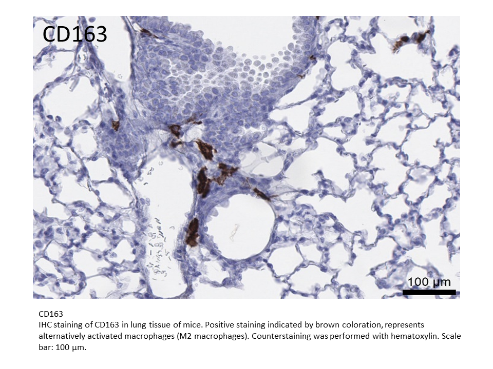
| CD163 | Ms, Hu, Rat | Membrane | 🔍Lung |
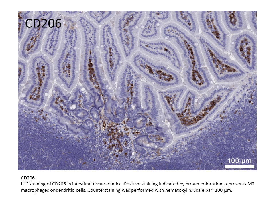
| CD206 | Ms | Membrane | 🔍Intestine |
| 神經科學(Neuroscience) | |||
抗體名稱 |
Reactivity |
染色部位 |
DEMO |
| Iba1 | Ms, Hu, Rat | Cytoplasm | 🔍Brain |
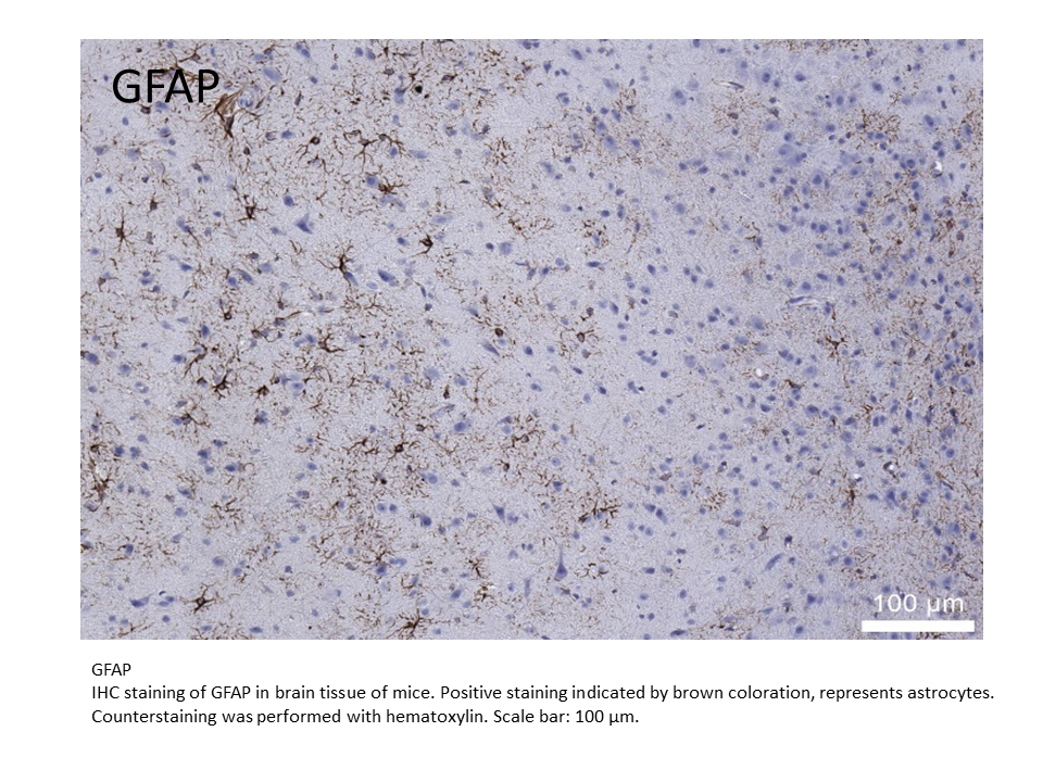
| GFAP | Ms, Hu, Rat | Cytoplasm | 🔍Brain |
| 代謝疾病/纖維化(Metabolism / Fibrosis / Oxidative Stress) | |||
抗體名稱 |
Reactivity |
染色部位 |
DEMO |
| α-SMA | Ms, Hu | Cytoplasm | 🔍Fibrotic liver |
| 4-HNE | - | Cytoplasm | |
| 標記用途/轉殖基因(Reporter or Tag-Based Detection) | |||
抗體名稱 |
Reactivity |
染色部位 |
DEMO |
| Luciferase | - | - | 🔍Tumor |
| GFP | - | - | 🔍Intestine |
| Ms, mouse; Rat, rat; Hu, human | |||