
Magnetic Resonance Imaging Facilities
Specific Aims
1. To provide MR resource and comprehensive technical support for research in magnetic resonance imaging and spectroscopy.
2. To conduct original research in new MR methods and foster interdisciplinary research into Medical Genomics.
3. To promote collaborative links between the MR and basic science research.
4. To provide education and training in the use of MR imaging technologies in biomedical research.
5. To link the pharmaceutical and biotechnology industries leading to potential partnerships in drug discovery and testing.
The specific MR techniques are intended to be set up for services:
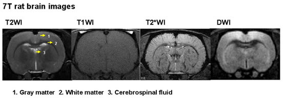
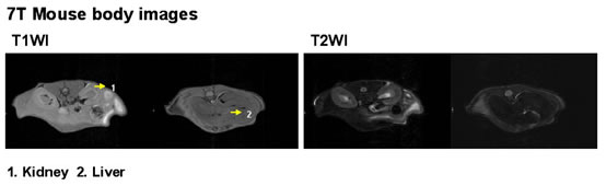
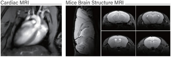
1. Standard T1-,T2-, and proton density weighted spin-echo and gradient echo imaging.
2. Contrast enhanced spin-echo imaging such as fat suppression, diffusion, magnetization transfer and fluid-attenuated inversion recovery.
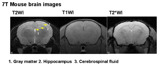
3. High-speed micro-anatomical standard and contrast enhanced spin-echo imaging at resolutions of about 30-50 microns.
4. In vivo/vitro high-resolution 3D isotropic volume imaging.
5. Assessment of tissue water diffusion, tissue-oxygenation and hemodynamics using fMRI methods including diffusion- and perfusion-weighted imaging and blood oxygenation level dependent imaging.
6. Mapping the white matter tracts using diffusion tensor imaging.
7. High-speed micro-anatomical diffusion-and diffusion tensor imaging.
8. Single-voxel and multi-voxel proton MR spectroscopy.
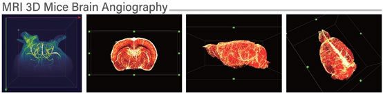
9. Magnetic Resonance Angiography.
10. Post-processing and analysis for displaying imaging and spectral data including volume determination, 3D volume rendering, spectral ratios, diffusion ratios, fiber tract mapping, and normalized cerebral flow (CBF) and volume (CBV) information.
The specific applications are intended to be set for services:
1. High-throughput phenotypic screens for neuro-and muscular-disorders.
2. Anatomical, functional and metabolic assessment of brain development and pathologies.
3. 3D white matter fiber tracking.
4. Assessment of the permeability of blood brain barrier (BBB).
5. Three-dimensional volume images from fixed specimens such as embryo and organ at an isotropic resolution of about 30-50 microns.
6. Monitoring both treatment progress and outcomes for in vivo assessment of prospective therapeutic drugs.
Supporting laboratories:
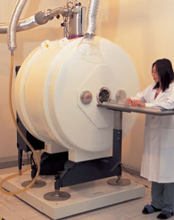
A. MRI Lab
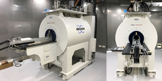
Bruker BioSpec 70/20 (National Biotechnology Research Park)
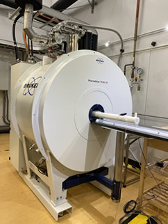
PharmaScan 70/16(Institute of Biomedical Sciences, Academia Sinica)
B. Post-Processing Lab
C. Immuno-Histology and Physiology Lab
D. Cell Culture Lab
Data Provided (For example):Please contact Dr. Yu-Wen Chen by dialing +886-2-2789-9027 to get more information.
Fees of Animal Imaging Facility (AIF) Services
Animal Imaging Facility (AIF) web